Frozen Fish-Men

If you know me, chances are you know of my love for the Hellboy comics by Mike Mignola. You’ve probably also heard me gush about how I met him and he signed a postcard for me and I got the postcard free and HE WAS JUST SO NICE. Ahem. Anyway, I’d been recommended that series for years but never listened to wisdom until I saw Ron Perlman punching Nazis on the big screen. I’ve been hooked ever since. This is all relevant because I recently bought one of the short story anthologies (Odder Jobs, in case you were curious) set in the Hellboy universe.  One of the stories involves Abe, the less tough, but equally awesome fish man that occasionally helps Hellboy and has adventures of his own. He’s also voiced by Niles from Frasier.

Image

Notice the resemblance? Neither do I.

So the relevant part is when the team is gathering to fight bad guys and everyone is bundled up against the cold except Abe, who seems surprised that everyone is so chilly. Hellboy has to point out that Abe is cold-blooded (ectothermic) and the rest of them are warm-blooded (endothermic). Except that isn’t correct. If Abe were just like an ectothermic lizard, he’d be even worse off in the cold. He’d be lethargic and miserable and useless for anything. If it were cold enough, he’d literally freeze; the water in his tissues would turn to ice because he can’t keep himself warm. However, Abe is a fish man, not a lizard man, so his body must have a strategy to prevent turning into a fish-sicle. Yes, I realize I’m questioning the biology of a fish-person, but it’s my blog and I do what I want! Work with me here, jeez.

There are actually a few strategies that ectotherms use to deal with dropping temperatures. For less extreme changes, such as between seasons or locations, ectotherms can adapt by restructuring membranes (to maintain proper fluidity even in the cold), regulating pH (to support enzyme function), adjusting enzyme concentrations and regulating isoforms (alternate forms of protein, with some better adapt to the cold than others). In cases of extreme cold, ectotherms have physiological methods to avoid or even tolerate freezing.

Some ectotherms that avoid freezing utilize organic osmolytes, carbon-based compounds that help regulate fluid balance. In this instance, the osmolytes are antifreeze proteins (often glyco-proteins, protein-sugar complexes), they lower the freezing point of bodily fluids, without damaging tissues. Thus they are called “compatible cryoprotectants”. Within the body and its cells, they cling on to and prevent the growth of ice. Other ectotherms can super-cool. This means that the body temperature will drop below freezing, but ice will not form because there is no trigger for it. However, external ice can act as a trigger for internal ice formation.

Ectotherms that are freeze tolerant do not avoid freezing at all. They will literally freeze solid, some animals will experience freezing of as much as 80% of their bodily fluids. The really miraculous thing is that they can survive this! CRAZY. They’re like winter zombies. A popular example of freeze tolerance is the spring peeper frog (which also has an ADORABLE name, I might add), which uses glucose as an antifreeze. Glucose floods the blood stream (450 times normal concentrations) and keeps the circulatory system pumping while the other 65% of the frog freezes. When the peeper thaws in the spring, the glucose doubles as quick energy

Now what does this all mean for Abe? Since he’s up and active in the winter and not frozen solid, it’s likely his body is filled with antifreeze proteins to keep active.

This also means Dark Horse Comics needs a better editor. Get it together, people!


Source

Sherwood, Lauralee, Hillar Klandorf and Paul Yancey. 2005. Animal Physiology: From Genes to Organisms. Thomson Brookes/Cole, Belmont, CA.

Yancey, Paul. “Subtidal Habitats- Polar Subtidal.” Marine Biology. Whitman College. Walla Walla, WA. 5 4 2011. Lecture.

Photo credit 2004 Hellboy movie, directed by Guillermo del Toro. Abe Sapien voice by Niles Crane David Hyde Pierce, with Doug Jones performing. Also photo credit from Frasier.

Sweet, Sweet Succes(ful Regulation of Blood Glucose)

I hope you all have been good little boys and girls and grown man/woman-children, because as promised I am going to explain blood glucose regulation. Bad children are temporarily banned from learning. Don’t even try to keep reading. You don’t want to know the punishment.

Anyway, first of all, take a look at this cake:

Image

LOOK AT IT.


We shall be considering this masterpiece of sweet, sweet heaven (courtesy of Stephanie Michaelis at Raspberri Cupcakes) for the rest of my post. Additionally, we have two scenarios: eat the cake and gaze longingly at photos of/not eating the cake.

Now, let’s imagine we get to eat some of this colorful, culinary delight. After some digestion, the pancreas recognizes the resulting increase in blood glucose. The pancreas has exocrine (secretes chemicals such as alkaline solution and digestive enzymes via ducts) and endocrine (secretes hormones directly into the bloodstream) tissues. Within the endocrine tissues are beta cells, the most abundant endocrine cell in the mammalian pancreas. Beta cells synthesize and secrete insulin in response to increased blood glucose. Insulin then travels to the liver and signals glycogenesis; the production of glycogen in skeletal muscle as well as the liver. Insulin will also inhibit gluconeogenesis (the production of glucose from molecules such as pyruvate or lactate) and facilitate glucose transport to cells. As a result, blood glucose will decrease to normal levels.

OK, back to reality. We do not get to eat the cake. We get to salivate with intense longing at the computer screen. We lose track of time. We forget to make a sandwich for lunch. Blood glucose drops. The pancreas detects this drop and other cells in its endocrine tissue, the alpha cells, produce and secrete glucagon into the bloodstream. Because the glucose…is GONE. Get it? Get it? Anyway, the liver receives this signal, glycogen production then decreases while glycogenolysis (glycogen break-down) increases along with gluconeogenesis. Blood glucose then increase to a more normal level.

Insulin and glucagon do not act exclusively of one another, with every spike and drop in blood glucose they work together to bring things back to normal. Of course, they can’t act instantly and can only go so far. For instance, Insulin would struggle if you inhaled an ENTIRE cake, while I can personally advise against long fasts if you want to do anything requiring focus like say…navigating Chicago by yourself on your first visit without passing out on the sidewalk on the way to the taxi. Those are bad life choices; spiking your blood glucose too high too often has long-term consequences and passing out on the sidewalk in a strange city has ALL the consequences.

So be street smart, carry snacks.


Source

Sherwood, Lauralee, Hillar Klandorf and Paul Yancey. 2005. Animal Physiology: From Genes to Organisms. Thomson Brookes/Cole, Belmont, CA.


Photo credit:

Michaelis, Stephanie. 2011. “Purple Ombre Sprinkles Cake.” Raspberri Cupcakes. 19 November, 2013 <http://www.raspberricupcakes.com/2011/11/purple-ombre-sprinkle-cake.html&gt;

Kidding Around Part 2

Back to goat guts.


Once a goat has masticated a mouthful (preferably grass and NOT my hair), it travels down the esophagus into the forestomach. The forestomach consists of the first three compartments of the entire stomach, the rumen, the reticulum and the omasum. At this point the food is less food and more of a wet mass. Scientists call it a bolus, but in ruminants it tends to be called cud. As in the stuff they regurgitate to chew on. Anyway, the rumen and reticulum are quite large, most of the anaerobic bacterial fermentation occurs here, besides some absorption of simple molecules and nutrients. Furthermore, food can loop from rumen, to reticulum back to the mouth a couple times before it is digested enough to move on to the omasum. Non-food items (like my hair) will make this trip a quite a few times, assuming the goat does not eventually give up (unlikely) or realize that this particular bite is not terribly appetizing (hella unlikely). Anyway, this loop of repeated mastication, fermentation and absorption greatly increases the efficiency of digestion. If you’ve ever wondered why it’s harder for humans to be herbivores, among other things, our omnivore digestive tract is a LOT less efficient at tackling an exclusively plant-based diet. Our digestive tract is more like that of a carnivore, particularly in terms of length (proteins are much easier to digest so carnivore digestive tracts and relatively short).

Again, back to the goat guts. When the bolus/cud has been chewed for the last time, it heads straight for the omasum. Here water and more nutrients are absorbed via the omasum’s highly folded surface. Oh, and get this: the omasum SORTS shit. Seriously. Big stuff that needs more ruminating will get chucked back (and possibly up-chucked) for more digesting. Anything not turned back continues on to the abomasum. The abomasum secretes acids to digest proteins and rumen microbes (remember the NPCs? They need to stay put or they’ll cause an infection) before the bolus moves on to the intestine as gooey chyme. Nummers. If the digesting goat in question is a kid, the abomasum will also secrete rennin (cheese, anyone?) to clot casein (milk protein) for better digestion.

With the high appetizing chyme now safely in the intestine, I think most of you get the rest: highly folded small intestine continues to absorb water and nutrients, chyme gets denser as it is moved via peristaltic waves (muscles squeezing it down the line) to the large intestine for compression and eventual expulsion. Probably right when the goat’s yard has just been swept.

You guys are lucky you’re so cute.

Image 


Source

Sherwood, Lauralee, Hillar Klandorf and Paul Yancey. 2005. Animal Physiology: From Genes to Organisms. Thomson Brookes/Cole, Belmont, CA.

Photo credit, me.

Kidding Around

OK, so goats.

Image

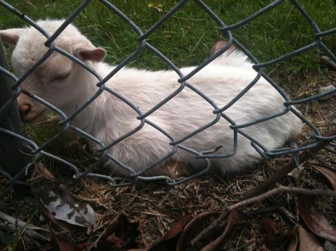
I love goats. Not just itty kiddies, but grown ones too. Not sure why, they’re not usually terribly cuddly. Although if you scritch that spot on their backs, right between the hind legs they just go weak in the knees and it’s adorable.

Anyway, in high school I volunteered at the local zoo’s (Woodland Park Zoo) “Family Farm” which featured goats and other farm animals that are part of the summer petting zoo. Besides tidying their stables, etc., we were responsible for keeping the goats and sheep social in the winter so that when summer came they did not flip any shits over small children trying to pet them. Or at least in one goat’s case, make sure he continued to ignore/avoid everything (he was not actually in contact area at all, but we continued to dream).

So we got used to the goaty quirks, like chewing on our hair and coats. You would also notice them stand still, not moving then suddenly belch softly and start chewing, mouth abruptly full. If you think they just regurgitated something to continue chewing on it…you are exactly right. Goats are herbivores, and considering the low nutritional value of plants, and the high difficulty in digesting large quantities of plant fibers, they possess a stomach with four compartments with which they tackle their dinners. The biggest contribution to digestion in goats comes from bacteria. Bacteria actually aid in fermentation of ingesta (ingested food/miscelani). Though periodically the goats need to regurgitate things that require more chewing.

That’s just a little introduction, but I will continue tomorrow.

Source

Sherwood, Lauralee, Hillar Klandorf and Paul Yancey. 2005. Animal Physiology: From Genes to Organisms. Thomson Brookes/Cole, Belmont, CA.

Photo credit, me.

Best of Buds 2

I know I promised more taste bud stuff yesterday, but I had spent the last part of work moving some VERY heavy equipment at work and I came home exhausted.

Image

Anyway, this will be short and sweet (sour, salty, bitter and savory). Despite what we all grew up learning about the “taste map” on our tongues, this is terrible and false. While taste buds will respond preferentially to certain flavors, they all respond at least a little to all flavors and there are no sour, salty, etc specific taste buds. Granted, there’s a little bit of mapping with where the taste buds with what preference sit on your tongue, but it varies from person to person.

Now! As to how these tastes actually get tasted, the answer…varies. But only by a little. Different channels and binding sites on the taste receptor cells respond to different molecules and ions to register flavors. Salty and sour flavors are registered when Na+ ions and H+ ions (respectively) come into contact with ion channels on the receptor cells. The resulting depolarization of the receptor cells causes it to signal the appropriate flavor. For sweet flavors, however, the sugar molecule (glucose, sucrose, artificial sugars can also work) attaches to a binding molecule on the receptor cell’s surface to cause cellular depolarization. Savory flavors involve tastants (taste molecules) binding to G-protein coupled receptors. I can talk about those cell surface receptors more later (if anyone even wants to know), but for now know they’re a kind of receptor protein. Anyway, savory flavors register when amino acids bind to the G-protein coupled receptor and cause cell depolarization. Bitter flavors, on the other hand, can be produced by a wide variety of molecules using a wide variety of binding/receptor mechanisms. Anything from alkaloids like caffeine and strychnine or metalloids like arsenic will register as bitter. Notice the inclusion of the poisons? Most animals register toxic substances as bitter, this is a survival strategy; we don’t like bitter things and thus avoid it. So if anyone gives you crap about putting sugar in your coffee, tell them you’re quieting your survival instincts. Also don’t judge, I do what I want!


Source

Sherwood, Lauralee, Hillar Klandorf and Paul Yancey. 2005. Animal Physiology: From Genes to Organisms. Thomson Brookes/Cole, Belmont, CA.

Photo credit me. Cheesecake credit, Kensie Steakhouse.

Best of Buds

Image

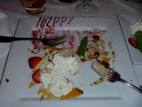
No, it is not my birthday, this is my cake from a birthday long (less than two years) past. Now, I am not a lady usually tempted by fancy cheesecake concoctions. Not really a lady in the first place. But if I saw pina colada or mint choco-brownie or raspberry-mocha-berry-fudgestravaganza cheesecake on a menu I’d typically reply with a scowl and a violent crossing of arms. So it is shocking I even considered hot buttered rum cheesecake. But consider it I did. Besides devouring and digesting it. This was not some weak cheesecake with delusions grandeur of being a dessert drink, this was hot buttered rum reincarnated as a cheesecake for good deeds in its previous life. It was glorious.

Image

My supreme enjoyment was all thanks to gustation. We all should thank our sense of taste; life would be a lot paler without the many tastes of our body fuel. Out would go birthday cake, fanciful cocktails, wine tasting and Cheezits. Spinach with bleu cheese salads, chocolate and mango juice would all be gone with the wind. We would just eat bizarre mashes of what’s good for us and enthusiastically tolerate it all. So then thank GOD AND ALL THAT IS HOLY for taste buds.

You have about 9000 taste buds and despite all the hype about them being color-coordinated taste microphones, they are a bit more complex than that. But just a bit. Taste buds are not the little nubbins all over your tongue, but the taste cells bordering the nubbins are the ones that really work the gustatory magic. The buds are composed of receptor cells and supporting cells. Receptor cells are modified epithelial (inner skin) cells that possess many folds to allow as much taste juice onto them as possible. This “tasting site” is the taste pore. Molecules and ions in the macerated food bind to the pore and results in a chain reaction: the receptor cells provoke an action potential (Remember action potentials?) in connecting nerve fibers and the brain in eventually alerted to the presence of tastiness (or not).

So what triggers this response? Well, it depends. Humans can taste sweet, salty, sour, bitter and meaty things and each have different ways to excite your senses. I will address this is detail tomorrow, but for now I will add that spicy is not a taste. It is your taste buds crying for a ceasefire.

Source

Sherwood, Lauralee, Hillar Klandorf and Paul Yancey. 2005. Animal Physiology: From Genes to Organisms. Thomson Brookes/Cole, Belmont, CA.

Photo credit me. Cheesecake credit, Kensie Steakhouse.

Get PUMPED

Hope you’re not tired of hearing my talking about blood and circulation, because surprise! Circulatory pathways! Specifically I want to show you how vertebrate circulation evolved from one simple circuit in early fish, to a dual circuit system in amphibians and mammals. To aid in explanation, I have provided my own diagrams (blame Medieval Musings).

Image

First, fish circulation. Fish kind of suck at circulation (hush up, ichthyologists, you know it’s true). Yes, their two-chambered heart suits an aquatic lifestyle and a more “sophisticated” three- or four-chambered might be a weight burden, but it’s basically a double-ended turkey baster. It needs auxiliary “hearts” (blood vessels with pumping capabilities) and constant forward motion to keep things moving in the right direction. So yeah, sucky circulation. However, it is at least easy to understand: deoxygenated blood gets pumped from the heart, to the gills where it is oxygenated, then to the body and finally back to the heart when the blood has been deoxygenated again.

Amphibian circulation is…fine…I mean, nothing wrong with it! They even have dual circuit circulation and a three-chambered heart to provide the pressure needed to pump blood when motionless. It’s just…muddled. Probably because when the oxygenated blood leaves the lungs and enters the heart, it mixes with deoxygenated blood before being pumped out to the body. So the blood that provides oxygen to the body isn’t fully loaded with the stuff. Like cars in the carpool lane, some of them are packed with people and some of them are packed with people-sized things. The liars.

The biggest difference between mammal hearts and amphibian hearts is just a little tissue. But that extra tissue means a four-chambered heart and fully oxygenated blood pumping from lungs to heart to body.

OK, that’s all for circulation…for now.

Image

Sources

-. 2010. GRE Subject Test: Biology 5th Ed. Kaplan, New York.

Sherwood, Lauralee, Hillar Klandorf and Paul Yancey. 2005. Animal Physiology: From Genes to Organisms. Thomson Brookes/Cole, Belmont, CA.

Oh boy! More Duvernoy!

So yesterday I gave y’all the idea that the Duvernoy secretes some sort of venom. Well…yes and no. Duvernoy secretions, like the gland itself, are highly variable and dependent on factors that may include age, diet and habitat (Chiszar and Smith 2002). These secretions have been classified as a venom for their mild to severe effects when released by the snake while feeding or defending (Kardong 2002). Pharmacological (drug) classification also identifies these secretions as venom, given their similarity to venom of “true” venomous snakes (Kardong 2002). Indeed, Duvernoy secretions have been seen to cause damage to the blood cells, nerves and muscles (Chiszar et al 2002; Weldon et al 2010). This is potentially related to pre-digestion of prey by snakes; the oral secretion of strong enzymes to degrade larger food items for easy consumption (Weldon et al 2010). However, these secretions lack certain enzymes that define typical venoms, possess an almost negligible fluid reservoir and have such a low release volume that they would be highly impractical as a true venom (Kardong 2002). Additionally, few muscles are associated with this gland to facilitate release, and much of its release is aided by chewing during feeding or defense (Chiszar and Smith 2002; Kardong 2002). This is supported by observations on the number of Colubrid bites on humans that do not result in envenomation, possibily due to short bite duration (Chiszar and Smith 2002). Defensive actions work best when it does not require hanging on so if Duvernoy secretions were meant to be used in defense, they would not require chewing motions to facilitate. Though this observation is scientifically unconfirmed, certain affects seen in snakebite victims seem more representative of allergic hypersensitivity reactions than symptoms of true envenomation (Chiszar and Smith 2002). With chemical analyses of Duvernoy’s gland secretions in abundance, behavior studies are lacking. Functional studies are yet needed to see how Colubrids actually use their “venom” (Kardong 2002).

Seems obvious, but it’s considerably harder to sit around with a camera and creep on snake lives than those of minor celebrities. It takes time and skill to obtain good results, so performing different chemical analyses that are more easily more accurate is very attractive for research.

Let me know if there are any questions, this was a bite from my thesis research, which I want to share but I get that it can be a bit dry.

Sources

Chiszar, D., and H. M. Smith. 2002. Colubrid Envenomations in the United States. Journal of Toxicology 21.1 & 2: 85-104.

Cundall, D., H. W. Greene. 2000. Feeding in Snakes. In: Schwenk, K. ed. Feeding: Form, Function, and Evolution in Tetrapod Vertebrates. San Diego: Academic Press.

Kardong, K. V. 2002. Colubrid Snakes and Duvernoy’s “Venom” Glands. Journal of Toxicology. 21.1 & 2: 1-19.

Kochva, E. 1979. Oral Glands of the Reptilia. In: Gans, C., A. Gans, eds, Physiology A, Vol. 5. London, New York: Academic Press.

Weldon, C. L., and S. P. Mackessy. 2010. Biological and Proteomic Analysis of Venom from the Puerto Rican Racer (Alsophis porticensis: Dipsadidae). Toxicon 55: 558-569.

Ahoy! It’s the Duvernoy!

It’s Sunday, it’s St. Patrick’s Day, let’s talk snake glands. Because that’s as close as I get to tangential. Specifically, I want to talk about the Duvernoy gland. I might even teach you how to pronounce it.

What is it? Homologue to the venom gland, the Duvernoy’s gland is probably the best studied of any colubrid head gland. By “homologue” I mean both the Duvernoy and the venom gland evolved from dental glands that began producing toxins and in early colubroids (subset of “modern snakes”)(Cundall and Green 2000). And by “colubrid”, I mean the group of snakes generally considered “non-venomous”. Besides having a suspiciously similar family name to the larger parent group Colubroidea, Colubridae (the group containing colubrid snakes) is pretty much a junk group. Taxonomists are like the rest of us; when they don’t know when to put/classify something, they have a scrap drawer to throw it in until they figure something out. Which means while it’s called the non-venomous group, there are actually no shared characters that define Colubridae. In fact, the highly venomous boomslang is a colubrid, it just doesn’t possess a venom gland or any other characteristics that would land it in a real family like Elapidae (cobras and friends) or Viperidae (guess). How are boomslang’s so venomous, then? Right! The Duvernoy.

Located over the upper jaw (Kochva 1979), the Duvernoy Gland is highly morphologically variable (Chiszar and Smith 2002) and typically associated with rear-fanged, semi-venomous snakes (Chiszar and Smith 2002). Semi-venomous snakes possess grooved (rather than tubular) fangs at the back of the mouth that release secretions at low pressure and their bite typically does not provoke strong reactions in humans (boomslang venom is one obvious exception)(Chiszar and Smith 2002; Kardong 2002). For the most part, Duvernoy glands are composed of protein-secreting (serous) cells, but they may also have mucous cells (Chiszar and Smith 2002). These cells are arranged in lobules that form ducts, as well as oral tissue that envelopes the base of the grooved fangs, which the Duvernoy ducts drain onto (Kardong 2002). The Duvernoy literally drains onto the fangs, so you don’t get a high pressure injection system as with traditionally venomous snakes. Colubrid snakes have to chew their prey a bit to work in the venom (Chiszar and Smith 2002). This could explain some of the variation found in colubrid bite severity in humans; severe bites could result from instances where the snakes got a good grip and started chewing (Chiszar and Smith 2002). Sounds fun, huh?

A little short tomorrow, but don’t you worry, there will be more on the Duvernoy tomorrow!


Sources

Chiszar, D., and H. M. Smith. 2002. Colubrid Envenomations in the United States. Journal of Toxicology 21.1 & 2: 85-104.

Cundall, D., H. W. Greene. 2000. Feeding in Snakes. In: Schwenk, K. ed. Feeding: Form, Function, and Evolution in Tetrapod Vertebrates. San Diego: Academic Press.

Kardong, K. V. 2002. Colubrid Snakes and Duvernoy’s “Venom” Glands. Journal of Toxicology. 21.1 & 2: 1-19.

Kochva, E. 1979. Oral Glands of the Reptilia. In: Gans, C., A. Gans, eds, Physiology A, Vol. 5. London, New York: Academic Press.

Weldon, C. L., and S. P. Mackessy. 2010. Biological and Proteomic Analysis of Venom from the Puerto Rican Racer (Alsophis porticensis: Dipsadidae). Toxicon 55: 558-569.