BAM. Thanks to the skilled investigations of a friend, we have a video of a rat snake in space/microgravity.
Enjoy.
BAM. Thanks to the skilled investigations of a friend, we have a video of a rat snake in space/microgravity.
Enjoy.

Because I can only be SO relevant to current events, today I am addressing homo…ology. Homology literally means “similar structures”, but in biology we differentiate between “serial” homology and “structural” homology. Both of these are very distinct from analogy.
First! Serial homology, for which snakes are an excellent example. Think of their ribs, you only have…uh, a limited number, but snakes have a lot more. Like the reticulated python above. Beautiful girl, 14 years old and 18 feet long (old enough and yet way too big for middle school. Poor thing will never get to prom), can you imagine the ribs she has? The many, many vertebrae and ribs of this python serve the same purpose in the same species and look…THE SAME. This is serial homology; those rib-vertebrae structures are in a series in the python and display homology.
Now, structural homology may require a slightly different image. Firstly, look at your hand, now look at the wings of this pooping Rodriguez Fruit Bat.

The bones in a bat wing are like elongate versions of your fingers. Just with some skin stretched between so that they can fly. Here, the structures are homologous, even if the function is not.
Finally, we have analogues. In biology, analogous structures have a similar function, but very different structures. Look at your hand, now look at this chicken.

NOW BACK TO YOUR HAND. Here are the fingers in the hand you love. THE FINGERS HAVE BECOME A BAT WING. By comparison the chicken wing is formed largely from only a couple fingers in the “hand” template, unlike the bat wing which uses all fingers. I’m on a horse. Anything is possible when you have functional structures with great flexibility in regards to adaptive variation.
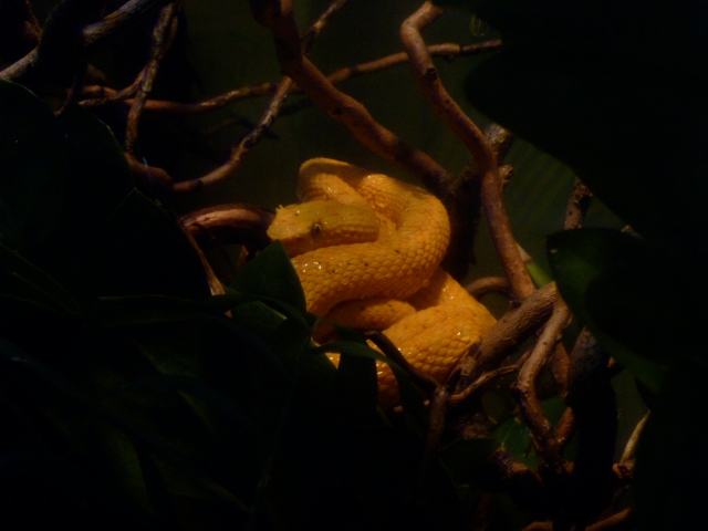
Eyelash Viper while I try to come up with a good topic for today.
So yesterday I gave y’all the idea that the Duvernoy secretes some sort of venom. Well…yes and no. Duvernoy secretions, like the gland itself, are highly variable and dependent on factors that may include age, diet and habitat (Chiszar and Smith 2002). These secretions have been classified as a venom for their mild to severe effects when released by the snake while feeding or defending (Kardong 2002). Pharmacological (drug) classification also identifies these secretions as venom, given their similarity to venom of “true” venomous snakes (Kardong 2002). Indeed, Duvernoy secretions have been seen to cause damage to the blood cells, nerves and muscles (Chiszar et al 2002; Weldon et al 2010). This is potentially related to pre-digestion of prey by snakes; the oral secretion of strong enzymes to degrade larger food items for easy consumption (Weldon et al 2010). However, these secretions lack certain enzymes that define typical venoms, possess an almost negligible fluid reservoir and have such a low release volume that they would be highly impractical as a true venom (Kardong 2002). Additionally, few muscles are associated with this gland to facilitate release, and much of its release is aided by chewing during feeding or defense (Chiszar and Smith 2002; Kardong 2002). This is supported by observations on the number of Colubrid bites on humans that do not result in envenomation, possibily due to short bite duration (Chiszar and Smith 2002). Defensive actions work best when it does not require hanging on so if Duvernoy secretions were meant to be used in defense, they would not require chewing motions to facilitate. Though this observation is scientifically unconfirmed, certain affects seen in snakebite victims seem more representative of allergic hypersensitivity reactions than symptoms of true envenomation (Chiszar and Smith 2002). With chemical analyses of Duvernoy’s gland secretions in abundance, behavior studies are lacking. Functional studies are yet needed to see how Colubrids actually use their “venom” (Kardong 2002).
Seems obvious, but it’s considerably harder to sit around with a camera and creep on snake lives than those of minor celebrities. It takes time and skill to obtain good results, so performing different chemical analyses that are more easily more accurate is very attractive for research.
Let me know if there are any questions, this was a bite from my thesis research, which I want to share but I get that it can be a bit dry.
Sources
Chiszar, D., and H. M. Smith. 2002. Colubrid Envenomations in the United States. Journal of Toxicology 21.1 & 2: 85-104.
Cundall, D., H. W. Greene. 2000. Feeding in Snakes. In: Schwenk, K. ed. Feeding: Form, Function, and Evolution in Tetrapod Vertebrates. San Diego: Academic Press.
Kardong, K. V. 2002. Colubrid Snakes and Duvernoy’s “Venom” Glands. Journal of Toxicology. 21.1 & 2: 1-19.
Kochva, E. 1979. Oral Glands of the Reptilia. In: Gans, C., A. Gans, eds, Physiology A, Vol. 5. London, New York: Academic Press.
Weldon, C. L., and S. P. Mackessy. 2010. Biological and Proteomic Analysis of Venom from the Puerto Rican Racer (Alsophis porticensis: Dipsadidae). Toxicon 55: 558-569.
It’s Sunday, it’s St. Patrick’s Day, let’s talk snake glands. Because that’s as close as I get to tangential. Specifically, I want to talk about the Duvernoy gland. I might even teach you how to pronounce it.
What is it? Homologue to the venom gland, the Duvernoy’s gland is probably the best studied of any colubrid head gland. By “homologue” I mean both the Duvernoy and the venom gland evolved from dental glands that began producing toxins and in early colubroids (subset of “modern snakes”)(Cundall and Green 2000). And by “colubrid”, I mean the group of snakes generally considered “non-venomous”. Besides having a suspiciously similar family name to the larger parent group Colubroidea, Colubridae (the group containing colubrid snakes) is pretty much a junk group. Taxonomists are like the rest of us; when they don’t know when to put/classify something, they have a scrap drawer to throw it in until they figure something out. Which means while it’s called the non-venomous group, there are actually no shared characters that define Colubridae. In fact, the highly venomous boomslang is a colubrid, it just doesn’t possess a venom gland or any other characteristics that would land it in a real family like Elapidae (cobras and friends) or Viperidae (guess). How are boomslang’s so venomous, then? Right! The Duvernoy.
Located over the upper jaw (Kochva 1979), the Duvernoy Gland is highly morphologically variable (Chiszar and Smith 2002) and typically associated with rear-fanged, semi-venomous snakes (Chiszar and Smith 2002). Semi-venomous snakes possess grooved (rather than tubular) fangs at the back of the mouth that release secretions at low pressure and their bite typically does not provoke strong reactions in humans (boomslang venom is one obvious exception)(Chiszar and Smith 2002; Kardong 2002). For the most part, Duvernoy glands are composed of protein-secreting (serous) cells, but they may also have mucous cells (Chiszar and Smith 2002). These cells are arranged in lobules that form ducts, as well as oral tissue that envelopes the base of the grooved fangs, which the Duvernoy ducts drain onto (Kardong 2002). The Duvernoy literally drains onto the fangs, so you don’t get a high pressure injection system as with traditionally venomous snakes. Colubrid snakes have to chew their prey a bit to work in the venom (Chiszar and Smith 2002). This could explain some of the variation found in colubrid bite severity in humans; severe bites could result from instances where the snakes got a good grip and started chewing (Chiszar and Smith 2002). Sounds fun, huh?
A little short tomorrow, but don’t you worry, there will be more on the Duvernoy tomorrow!
Sources
Chiszar, D., and H. M. Smith. 2002. Colubrid Envenomations in the United States. Journal of Toxicology 21.1 & 2: 85-104.
Cundall, D., H. W. Greene. 2000. Feeding in Snakes. In: Schwenk, K. ed. Feeding: Form, Function, and Evolution in Tetrapod Vertebrates. San Diego: Academic Press.
Kardong, K. V. 2002. Colubrid Snakes and Duvernoy’s “Venom” Glands. Journal of Toxicology. 21.1 & 2: 1-19.
Kochva, E. 1979. Oral Glands of the Reptilia. In: Gans, C., A. Gans, eds, Physiology A, Vol. 5. London, New York: Academic Press.
Weldon, C. L., and S. P. Mackessy. 2010. Biological and Proteomic Analysis of Venom from the Puerto Rican Racer (Alsophis porticensis: Dipsadidae). Toxicon 55: 558-569.
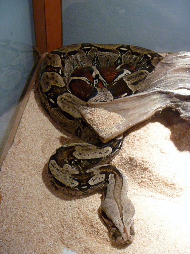
I keep looking for a reason to post a picture of this snake, but none have come up so here it is. This is a red-tailed boa.
Enjoy.
